Shou-Liang Wang1,
Lian Duan2,
Wei Li1,
Gong-Ming Wang1,
Meng-Yuan Zhang1 ![]()
For correspondence:- Meng-Yuan Zhang Email: zhangmy1976@gmail.com Tel:+8653168776472
Received: 10 January 2016 Accepted: 30 September 2016 Published: 25 February 2017
Citation: Wang S, Duan L, Li W, Wang G, Zhang M. Genistein attenuates ischemia/reperfusion injury in rat kidneys via enhancement of antioxidant defense mechanisms: Activation of Nrf-2/HO-1 signaling. Trop J Pharm Res 2017; 16(2):349-356 doi: 10.4314/tjpr.v16i2.13
© 2017 The authors.
This is an Open Access article that uses a funding model which does not charge readers or their institutions for access and distributed under the terms of the Creative Commons Attribution License (http://creativecommons.org/licenses/by/4.0) and the Budapest Open Access Initiative (http://www.budapestopenaccessinitiative.org/read), which permit unrestricted use, distribution, and reproduction in any medium, provided the original work is properly credited..
Purpose: To investigate the protective role of genistein against ischemic reperfusion (I/R) injury in rat kidneys.
Methods: Group I (control, n = 10) consisted of animals that were not operated on while group II (sham, n = 10) were animals surgically operated on, similar to I/R group without renal bilateral ischemia. Group III (genistein, n = 10) consisted of animals administered 10 mg/kg genistein by oral gavage for 7 consecutive days while group IV (I/R, n = 10) animals were subjected to 45 min of renal bilateral ischemia followed by 24 h of reperfusion. Group V (genistein+I/R, n = 10) received 10 mg/kg genistein by oral gavage for 7 consecutive days and then subjected to 45 min of renal bilateral ischemia followed by 24 h of reperfusion. Renal function, total oxidant capacity and total antioxidant status in serum were evaluated in the rats. Further, reactive oxygen species generation as well as levels of protein carbonyl, lipid peroxidation, and enzymatic and non-enzymatic antioxidants were determined. Nrf-2 (nuclear factor (erythroid-derived 2)-like 2) and HO-1 (Heme oxygenase-1) ex
Results: Pre-treatment with genistein (10 mg/kg) significantly (p < 0.001) ameliorated I/R induced renal damage by reducing the levels of serum markers. Genistein pre-treatment significantly decreased (p < 0.001) I/R injury induced-ROS, lipid peroxides and protein carbonyl content (p < 0.001). I/R injury significantly (p < 0.001) decreased non-enzymatic and enzymatic antioxidant activities. Genistein pre-treatment also prevented renal I/R injury by significantly up-regulating Nrf-2, HO-1 ex
Conclusion: Thus, genistein may be therapeutically useful against kidney I/R injury by improving antioxidant defense mechanisms.
Introduction
Renal ischemia/reperfusion injury (IRI) causes kidney dysfunction with increased mortality rate [1]. Oxygen impairment and subsequent accumulation of waste products initiates tissue damage [1,2]. Reperfusion causes increased free radical production ultimately leading to cellular death through apoptosis/necrosis [3]. Oxidative stress and decline in antioxidant balance are central regulatory mechanism in the pathogenesis of renal I/R injury [4]. Pre-conditioning offers protective effect against I/R injury and pre-treatment with pharmacological agents might be a possible therapeutic option against renal I/R injury [5].
Genistein, a plant derived isoflavone has potent anti-oxidative property and offers numerous health benefits including cardioprotection, anti-inflammatory and anti-cancer effects [6,7]. In the present study, we aimed at investigating whether genistein could offer protection against renal ischemic reperfusion injury by regulating oxidative stress and Nrf-2 signaling.
Methods
Animals and study design
All the animals and protocols used in the present study were followed according to National Research Council for Animal Care [8] and approved by the Ethical Committee (no. NR 016/2014) of Shandong Provincial Hospital affiliated to Shandong University, China, prior to the commencement of the study. Fifty Male Wistar rats (age of 8 - 10 weeks) weighing 180 - 220 g housed under standard conditions. The rats were maintained under controlled conditions of (21 ± 2 ºC) and relative humidity (65 - 70 %) with alternating 12 h dark and light cycle. The animals were provided with ad libitum standard laboratory diet and water. After acclimatization, animals were divided randomly into 5 groups with ten animals per group. Genistein and all other chemicals used in the study were purchased from Sigma Aldrich (Shanghai, China).
Study design
Group I (control, n = 10) consisted of animals that were not operated on while group II (sham, n = 10) were animals surgically operated on, similar to I/R group without renal bilateral ischemia. This was followed by midline laparotomy; the kidneys were located and the renal pedicles with artery, vein and nerve supply were subjected to bilateral ischemia for 45 min. Group III (genistein, n = 10) consisted of animals administered 10 mg/kg genistein by oral gavage for 7 consecutive days while group IV (I/R, n = 10) animals were subjected to 45 min of renal bilateral ischemia followed by 24 h of reperfusion. Group V (genistein+I/R, n = 10) received 10 mg/kg genistein by oral gavage for 7 consecutive days and then subjected to 45 min of renal bilateral ischemia followed by 24 h of reperfusion. At the end of treatment period, the animals were fasted for 12 h, and anaesthetized with xylazine (10 mg/kg) and ketamine (100 mg/kg). Heating lamp was used to maintain body temperature (37.5 ºC). Abdominal region was sterilized with povidine iodine solution and both the kidneys were isolated by midline incision procedure. The abdomen was closed in the sham group alone. In I/R and genistein + I/R administered groups, non-traumatic microvascular clamps were used to occlude renal arteries for 45 min. After 45 min, the clamps were removed blood flow was restored to the kidney and the incision closed.
After 24 h reperfusion, the animals were anesthetized and blood was collected by cardiac puncture and the separated serum used for serum biomarker analysis. Nephrectomy was performed and the isolated kidney used for biochemical and molecular studies.
Assessment of renal function markers
Serum urea nitrogen was determined as described previously [9]. To 0.1 ml of serum, 3.3 ml of double-distilled water, 0.3 ml of 10 % sodium tungstate and 0.3 ml of 0.67 N sulfuric acid were added. The samples were centrifuged at 4000 rpm, 15 min). To the supernatant, 1 ml of double-distilled water, 0.4 ml of diacetyl monoxime reagent and 0.6 ml of sulfuric acid-phosphoric acid mixture were added and in a boiling water bath for 30 min. The samples were cooled to room temperature and the absorbance was read at 480 nm in a Shimadzu UV - 1700 spectrophotometer. Creatinine was estimated using the method described previously [10]. To 1.0 ml of serum 8.0 ml of double-distilled water, 0.5 ml of 2/3 N sulfuric acid and 0.5 ml of 10 % sodium tungstate were added and centrifuged (4000 rpm, 15 min). To 5.0 ml of the clear supernatant, 1.5 ml of saturated picric acid and 1.5 ml of 0.75 N sodium hydroxide were added and incubated for 15 mins. The absorbance was read at 460 nm in Shimadzu UV - 1700 spectrophotometer.
Evaluation of serum total antioxidant capacity (TAC) and total oxidant status (TOS)
Serum TAC levels were determined using a novel automated measurement method [11]. The hydroxyl radical produced through free radical reactions is directly proportional to the antioxidant levels in the serum. The results are expressed as μmol Trolox Eq/L. Serum TOS values were determined as described previously [12]. The colour intensity is directly proportional to the oxidant molecules in the serum. The assay is calibrated with hydrogen peroxide (H2O2) and the results are expressed in terms of micromolar hydrogen peroxide equivalent per litre (μmol H2O2 Eq/L).
Measurement of oxidative stress and antioxidant status
The tissues were homogenized using ice-cold 50 mM Tris-HCl buffer (pH 7.4) and centrifuged at 3,000 rpm for 15 min (KUBOTA, KR-20000T). The supernatant was aliquoted and stored at - 20 ºC for further analysis. Protein estimation was performed as described previously [13].
Determination of reactive oxygen species
The levels ROS was determined as described previously [14]. To the kidney homogenate added 5 mM potassium phosphate buffer (pH 7.4) and 2',7' -dichlorofluorescein diacetate DCF-DA (10 µM), incubated at 37 ºC for 15 min followed by centrifugation (10,000 rpm, 10 min). The pellet suspended in phosphate buffered saline was incubated for 60 min at 37 ºC. Fluorescence intensity was measured at excitation (485 nm) and emission (528 nm) in a spectrofluorometer (Thermo Scientific, Waltham, MA). The results were expressed in percentage ROS generation was computed relative to control value and expressed as percentage.
Determination of tissue protein carbonyl content (PCC)
Tissue protein carbonyl content was determined as described previously [15]. The samples were treated with 10 mM DNPH in 2 M HCl and incubated at room temperature for 1 hr (shaking). The hydrazone derivatives were precipitated with 20 % trichloroacetic acid and then was treated with ethanol/ethyl acetate, 1:1 to remove excess DNPH, and then re-precipitated with 10 % trichloroacetic acid. The pellet was dissolved in 6 M guanidine hydrochloride. The absorbance was measured at 370 nm (Shimadzu UV - 1700 spectrophotometer). The results were calculated as nmole of DNPH incorporated/mg of protein. The results were expressed as nanomoles of carbonyl/mg of protein.
Lipid peroxidation assay
Thiobarbituric acid reactive species (TBARS) content was determined as described previously [16]. To the 0.1 ml homogenate added, 200 μl of butylated hydroxytoluene (BHT) in methanol, 200 μl 1M phosphoric acid and 350 μl of TBA (Thiobarbituric acid) solution. The tubes were mixed vigorously and incubated for 60 min at 60 °C. The mixture was centrifuged at 10,000g for 10 mins. The supernatant was read at 532 nm. The amount of TBARS formed was calculated using extinction coefficient, E = 1.56 x 10⁵). The results are expressed as thiobarbituric acid reactants formed/mg of protein.
Evaluation of antioxidant enzyme activities
Glutathione (GSH)
The samples were precipitated with using TCA and centrifuged at 6000 g for 15 mins. The supernatant was treated with DTNB (5,5′-dithiobis- (2-nitrobenzoic acid), where the –SH group of GSH reacts with DTNB and the yellow coloured product was measured at 405 nm. The GSH content was determined using the standard GSH concentration. The results were expressed as Units/mg of protein. 1U = nmoles of GSH/mg of protein.
Superoxide dismutase (SOD) activity
The SOD activity was determined as described [17]. To 0.1 ml of homogenate, added 50 mM Tris-cacodylic acid buffer (pH 8.2), and pyrogallol solution (0.2 mM) and EDTA (1 mM). Followed by which the reading was measured for 3 mins (30 sec interval). The method measures the auto-oxidation of pyrogallol and 1 unit = the amount of enzyme required to inhibit pyrogallol auto-oxidation by 50 %.
Glutathione-S-Transferase (GST) activity
The reaction mixture contained 0.1 ml of homogenate in 0.1 M phosphate buffer (pH 6.5), 1 mM 1-chloro-2, 4-dinitrobenzene (CDNB), and 1 mM GSH in a final volume of 3 ml. Followed by which the reading was measured for 3 mins (30 sec interval). The enzyme activity was determined using extinction coefficient of GS-CDNB, E340 = 0.0096 μM− 1 cm− 1 and expressed as Units/mg of protein. 1U = nmoles of CDNB conjugated/min/mg protein [18].
Glutathione peroxidase (GPx) activity
GPx was performed as described by [19]. To 0.1 ml of homogenate in 50 mM phosphate buffer pH 7.0, added 0.5 ml sodium azide (0.1 mM), 1ml glutathione (0.5 mM), 1.2 ml hydrogen peroxide (1 mM). To the above reaction mixture added 0.5 ml of 10 % TCA and incubated for 5 mins at 37 °C and centrifuged at 5000 rpm for 15 mins. To the supernatant (0.1ml), added 5,5′-Dithio-bis (2-nitrobenzoic acid) (DTNB) and the read at 412 nm. 1U = 1 μ mole GSH to GSSG in the presence of H2O2/min. The enzyme activity was expressed as units/mg protein.
Catalase (CAT) activity
The activity was determined according to the method described by [20]. To 0.1 ml of tissue homogenate added, 50 mM, phosphate buffer, pH 7.0. 1.0 ml of 10 mM H2O2 was added and the reading was measured for 3 mins (30 sec interval) at 240 nm. The enzyme activity was determined using extinction coefficient of H2O2 (E240 = 43.6 M− 1 cm− 1) and expressed as Units/mg of protein. 1U = μM of H2O2 utilized/min/mg protein.
Western blot analysis
The tissues were homogenized and nuclear and cytoplasmic extract were isolated from CelLytic Nuclear Extraction kit. The protein isolated was loaded in 12 % SDS-PAGE gels and the separated proteins were transferred into NC (nitrocellulose) membrane. The membrane was incubated with primary and secondary antibodies for Nrf-2 and HO-1 proteins and developed with super signal ECL kit. The bands were developed and analyzed using Image J software.
Statistical analysis
The data were analyzed using one-way analysis of variance (ANOVA) followed by Tukey’s multiple comparison test using SPSS version 10.0). The level of significance was set variously at p < 0.05, p < 0.01 and p < 0.001.
Results
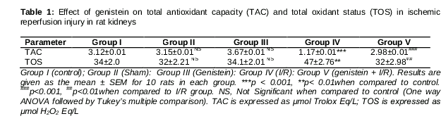
Renal I/R injury caused significant increase (p < 0.001) in serum kidney markers urea and creatinine when compared to control rats. Sham rats and Genistein treated rats showed non-significant difference in serum biomarkers when compared to control group. Treatment with Genistein followed by I/R restored the kidney function by reducing renal markers (p < 0.001) compared to I/R rats (Fig1A and 1B). I/R rats showed significant increase (p<0.001) in TOS levels with concomitant decrease (p < 0.01) in TAC levels compared to control group. However, in rats pre-treated with genistein followed by I/R injury showed significant decrease (p < 0.001) in TOS levels with concomitant increase (p < 0.01) in TAC ().
Genistein ameliorates I/R injury
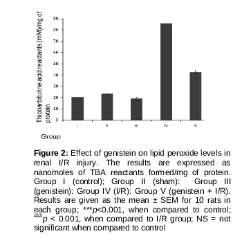
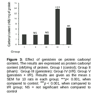
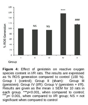
Rats with I/R injury showed a significant increase (p < 0.001) in the levels of lipid peroxides (), protein carbonyl content () and reactive oxygen species () when compared to control rats. These levels were significantly decreased (p < 0.001) in rats treated with genistein followed by I/R injury. Rats treated with genistein/sham-operated rats showed non-significant levels of oxidative markers when compared to control group.
Genistein prevents renal I/R injury
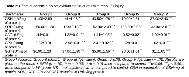
The levels of GSH found to be significantly decreased (p < 0.001) in I/R rats when compared to control group. Genistein treatment increased the GSH levels in a significant manner (p < 0.001) when compared to I/R alone treated rats (). The activities of enzymatic antioxidants (SOD, CAT, GST and GPx) were significantly declined when compared to control group. These antioxidant enzyme activities were significantly increased in rats treated with genistein followed by I/R injury. There was a non-significant difference in the antioxidant activities in sham rats and Genistein treated rats when compared to control group ().
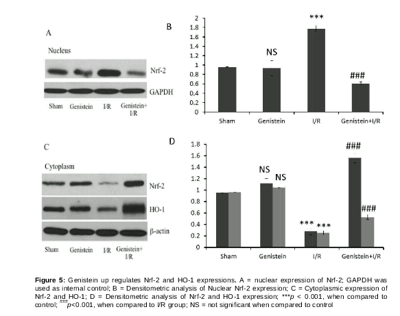
Genistein pre-treatment induces Nrf-2/HO-1 expression during I/R injury
In order to determine whether protective effect of genistein is mediated by nrf-2 signaling we analyzed the expressions Nrf-2 and HO-1 in the wistar rats. shows that I/R injury caused significant decrease of Nrf-2 levels in the nucleus however treatment with genistein followed by I/R injury increased nuclear Nrf-2 expressions. Further, HO-1 expression was significantly down regulated during I/R injury, while pre-treatment with genistein increased the HO-1 expressions compared to I/R injury group.
Discussion
The present study shows that genistein significantly offered cytoprotection against renal ischemic reperfusion injury in wistar rats. The increased level of serum markers (urea and creatinine) during I/R injury demonstrates severe damage to the renal tissue. These levels were markedly reduced during genistein treatment in rats with I/R injury. Further, genistein exerted protective effect by increasing the antioxidant capacity with concomitant decline in total oxidant status. Similar protective effect offered by genistein against renal damage by decreasing the serum urea and creatinine levels has been reported earlier [21].
Over production of ROS leads to oxidative stress conditions, which ultimately results in tissue injury. One of the most sensitive organs for oxidative stress includes kidney and the present study showed a significant increase in the levels of reactive oxygen species during renal I/R injury. Intracellular reactive oxygen species include species such as hydrogen peroxide (H2O2), hypochlorous acid (HClO), superoxide anion (O2-). Any imbalance in these levels leads to rise in ROS levels, which results in damage to DNA, lipid and proteins. Reactive species interact with unsaturated bonds in lipid moieties and leads to the formation of lipid peroxides [22]. These species also result in oxidation of proteins and ultimately results in the formation of protein carbonyls. The stable moieties are measured biochemically, which acts as an important biomarker in protein damage [23]. The present studies showed a significant increase in lipid peroxide levels and protein carbonyl content during I/R injury. A significant accumulation of MDA and protein carbonyls during I/R injury was ameliorated during genistein treatment. Additionally, Genistein treatment during ischemic reperfusion injury reduced reactive oxygen species generation, thus it might be the reason for its preventive effects against various downstream events against I/R induced oxidative stress. The antioxidant property of genistein against the amelioration oxidative stress status through inhibition of lipid peroxidation and ROS generation has been documented earlier [24and Guidry 2000].
It has been well documented that antioxidant defense mechanisms play a crucial role in protecting against various diseased conditions, which is primarily mediated through oxidative stress. Various phase II cytoprotective enzymes such as SOD, CAT, GST and GPx are involved in protective role. GSH is an important non-protein thiol, which is endogenous in nature, acts as a first defense against oxidative stress conditions [25]. Enzymatic antioxidants act in coherence in the efficient removal of toxic species from the cell. Superoxide dismutase reacts with superoxides and results in the formation of hydroxyl radicals. Catalase reacts with the hydroxyl radicals and forms H2O and O2 [26]. GSH in conjugation with GPx offers protection from various toxic metabolites [27]. The present study shows that I/R injury lead to decrease in levels of GSH content and enzymatic antioxidants (superoxide dismutase, catalase, glutathione peroxidase and glutathione-S-transferase). However, genistein treatment followed by I/R injury showed a significant increase in both enzymatic and non-enzymatic antioxidant status. Further, we also demonstrate here that Genistein induced protection against I/R injury was mediated through Nrf-2/HO-1 signaling mechanisms. Nrf-2 (nuclear factor erythroid 2-related factor 2) is a nuclear transcription factor and well established as oxidative stress regulator. Under normal and oxidative stress conditions, Nrf-2 controls the antioxidant defense mechanisms. Nrf-2 translocates into nucleus and binds to antioxidant response elements (ARE)- and induces expression of HO-1, GCL, NQO1 [28]. Genistein induced improved antioxidant enzyme activities have been reported earlier and the mechanism of protection has been linked with up-regulation of Nrf-2 activation and expression [29].
Conclusion
The findings of the present study show clear evidence that genistein treatment offers significant protective effect against renal ischemic reperfusion injury in rats by reducing renal damage and oxidative stress with concomitant increase in antioxdative defense mechanisms. Thus, nutritional intervention with genistein may be a promising strategy against ischemic reperfusion injury.
References
Archives


News Updates